
Hair Transplant Microscope: How It Works, Cost, Results
What a hair transplant microscope is, how surgeons use it to place grafts, recovery, results, who it suits, and cost by stage of hair loss.
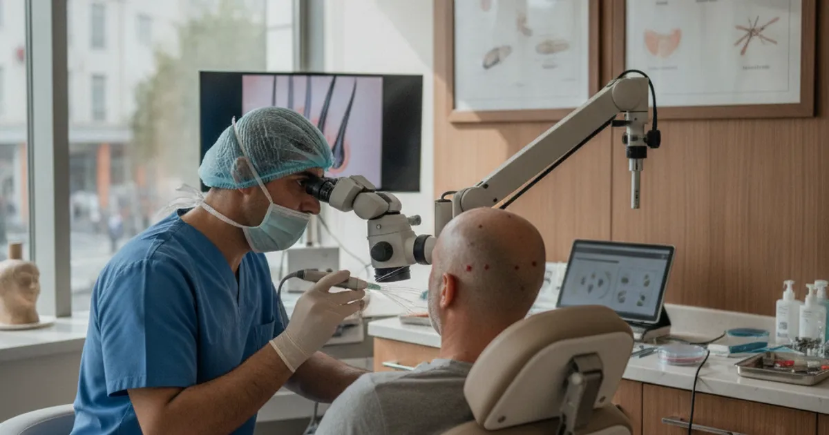
What Is a Hair Transplant Microscope?
A hair transplant microscope is exactly what it sounds like: a high-magnification optical instrument a surgical team uses to see individual hair follicles and the tiny grafts cut from a strip of scalp. In a follicular unit transplantation (FUT) procedure, the surgeon removes a strip of donor scalp, and the team then slices that strip into grafts containing one, two, three, or four hairs. Doing that by eye is possible, but it is rough. Doing it under a stereoscopic microscope lets the team separate grafts cleanly, keep the follicles intact, and sort them by hair count before implantation. The microscope is not a gimmick or a brand name. It is a piece of surgical equipment, and whether a clinic uses one tells you something about how carefully they handle your donor hair.
- Magnifies the donor strip so technicians can see individual follicular units and the surrounding tissue
- Helps avoid transecting (slicing through) follicles, which kills the graft
- Allows grafts to be sorted into 1-hair, 2-hair, 3-hair and 4-hair groups for precise placement
- Reduces the time grafts sit outside the body in a dehydrated state
- Commonly paired with backlighting and chilled holding solutions to keep grafts viable
How Microscopes Are Used During a Hair Transplant
The microscope comes into play in the middle of the procedure, after the donor strip has been removed and before the grafts go into the recipient area. Here is the general sequence in a microscope-assisted FUT case.
Graft dissection under a stereoscopic microscope is where the microscope earns its keep.
Types of Microscopes Used in Hair Restoration
Not all microscopes are the same, and clinics use different setups depending on their technique and budget. The two main categories are stereoscopic (dissecting) microscopes and the older single-lens variety. Some clinics also use video-microscope systems that project the magnified image onto a screen so the whole team can see what is happening.
| Microscope Type | How It Works | Typical Use |
|---|---|---|
| Stereoscopic (dissecting) microscope | Two eyepieces give a 3D view; magnification typically 5x to 25x | Standard for graft dissection in FUT |
| Single-lens microscope | One eyepiece, flat 2D image; cheaper but less depth perception | Older setups, smaller clinics |
| Video microscope | Camera projects magnified image onto a monitor | Teaching, team coordination, documentation |
| Surgical loupes | Worn on the head; 2x to 6x magnification | Recipient site creation and implantation, not dissection |
- Stereoscopic microscopes are the gold standard for graft dissection because the 3D view helps technicians avoid cutting follicles.
- Loupes are not a substitute for a microscope during dissection, though some clinics use them for implantation.
- A clinic that dissects under a microscope is generally handling grafts more carefully than one that does not.
- Ask which magnification range the clinic uses; higher is not always better, but 5x to 10x is typical for clean dissection.
Recovery and Results with Microscope-Assisted Transplants
Recovery from a microscope-assisted transplant is not dramatically different from any other FUT procedure. The microscope affects graft survival and hair yield, not how fast your scalp heals. You will still have a sutured donor line, scabbing in the recipient area, and a few weeks of redness. The main difference shows up months later, when the grafts start growing.
- Days 1 to 3: mild swelling, scabbing, and tenderness in both donor and recipient areas
- Days 4 to 10: scabs fall away, sutures or staples come out around day 10 to 14
- Weeks 2 to 8: transplanted hairs shed as part of the normal cycle; this is expected
- Months 3 to 6: new growth begins, initially fine and uneven
- Months 9 to 12: most of the final result is visible; density continues to mature up to 18 months
Who Is a Good Candidate?
Microscope-assisted surgery is not a separate candidacy category. If you are a candidate for a hair transplant at all, you are a candidate for one done under a microscope. That said, some situations benefit more than others.
- People with a stable donor area and enough density to support a strip harvest
- Those wanting maximum graft yield from a limited donor supply
- Patients with fine or curly hair, where grafts are harder to dissect cleanly by eye
- Anyone planning a large session where graft handling time is longer
- People who want the option of FUT rather than FUE, since the microscope is most associated with strip dissection
Cost of Microscope-Assisted Hair Transplants
Pricing for a microscope-assisted transplant is not usually listed as a separate line item. Clinics that use microscopes tend to price by graft count or by procedure, and the microscope is part of their standard setup. What matters more for your budget is how advanced your hair loss is, because that determines how many grafts you need. Below are averaged case prices by stage of hair loss.
| Stage of Hair Loss | Typical Graft Range | What Drives the Price |
|---|---|---|
| Early | 1,000 to 1,500 grafts | Smaller session, shorter surgery time |
| Moderate | 1,500 to 2,500 grafts | More dissection and implantation time |
| Advanced | 2,500 to 4,000+ grafts | Large session, longer graft handling, more staff |
Because microscope-assisted dissection is labour-intensive, clinics that use it often charge at the higher end of the local market. That is not a reason to avoid it, but it is a reason to compare quotes carefully and ask what is included. Use the directory to compare clinics and see what each one offers.
How It Compares to Other Techniques
The microscope is not a competing technique to FUT or FUE. It is a tool used within those techniques, mostly during the dissection stage. Here is how it stacks up against the alternatives.
| Approach | Magnification Used | Graft Handling | Best For |
|---|---|---|---|
| Microscope-assisted FUT | Stereoscopic microscope, 5x to 25x | Very precise dissection, sorted grafts | Maximising yield from a strip |
| Loupe-assisted FUT | Surgical loupes, 2x to 6x | Less precise, faster | Smaller sessions, lower cost |
| FUE with loupes | Loupes, 2x to 6x | Individual extraction, no strip | Avoiding a linear scar |
| FUE with microscope | Microscope for graft cleaning | Precise cleaning and sorting | Large FUE sessions, fine hair |
- Microscope-assisted FUT generally gives the highest graft yield per strip because fewer follicles are damaged.
- FUE does not always use a microscope, but some clinics use one to clean and sort extracted grafts.
- Loupe-only dissection is faster and cheaper but carries a higher risk of transection.
- The right choice depends on your donor density, hair type, and whether you want to avoid a linear scar.
Choosing a Clinic That Uses Microscopes
If you want a microscope-assisted procedure, you need to ask directly. Not every clinic advertises it, and some use the term loosely. Here is what to look for.
- Ask whether graft dissection is done under a stereoscopic microscope or with loupes only.
- Ask how many technicians work at microscopes and how many grafts each handles per session.
- Ask about graft holding solution and chilling, since these matter as much as magnification.
- Ask for the clinic's graft survival rate or how they measure it.
- Check whether the surgeon or technicians do the dissection, and how long they have been doing it.
Frequently Asked Questions
Is a microscope-assisted hair transplant better than one done with loupes?
For graft dissection, yes, in most cases. A stereoscopic microscope gives a 3D magnified view that makes it easier to separate follicular units without cutting through them. Loupes are faster and cheaper but carry a higher risk of transection. The difference shows up in graft yield, not in how the procedure feels.
Does using a microscope change recovery time?
No. Recovery is determined by the surgical technique (FUT or FUE) and your own healing, not by the magnification used during dissection. You will still have scabbing, redness, and a shedding phase in the first few months.
Is a microscope-assisted transplant more expensive?
It can be, because dissection under a microscope is slower and requires more skilled technicians. But clinics rarely price it as a separate add-on. Compare total quotes by graft count and stage of hair loss rather than assuming a microscope automatically means a higher bill.
Can I get an FUE transplant with a microscope?
Yes. Some clinics use a microscope to clean and sort grafts after FUE extraction, even though the extraction itself is done with a punch and loupes. It is less common than microscope-assisted FUT but not unusual.
More Guides
- Hair Transplant for Curly Hair: Graft and Technique Tips
A plain-English guide to hair transplants for curly hair: how surgeons adapt graft harvesting and placement, recovery, results, costs, and who it suits.
- How Long Does a Hair Transplant Last? Long-Term Durability
How long does a hair transplant last? We explain the procedure, recovery, durability, costs by stage of hair loss, and how it compares to alternatives.
- Hair Transplant Revision: When a Second Procedure Makes Sense
Considering a hair transplant revision? Learn how a second procedure works, who it suits, recovery, results, and costs. Expert explainer from Clinic Reccy.
- Hair Transplant for Norwood 6 and 7: What's Possible
A clear look at hair transplant options for Norwood 6 and 7 hair loss: graft limits, staging, recovery, costs, and realistic results.